Toggle
contrast
mode
X-ray imaging
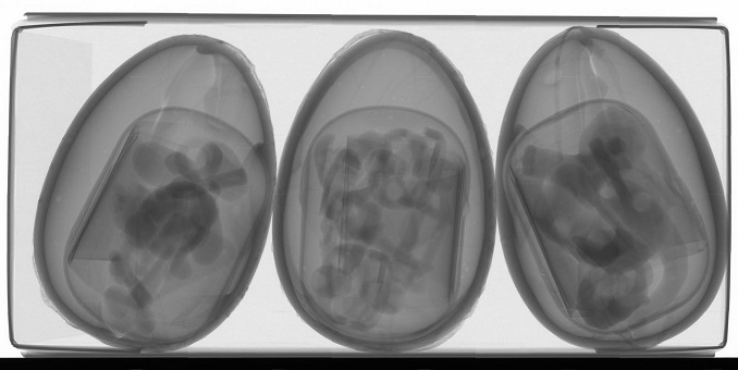
The ability of X-rays to pass through opaque objects makes it possible to see the inside of all kinds of samples. The image below is an X-ray image of a paper carton containing three chocolate eggs.

X-ray phase contrast
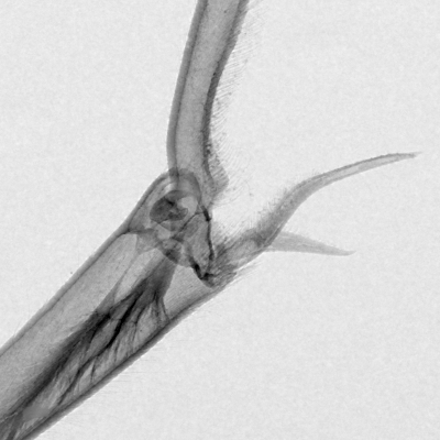
X-ray phase contrast is a set of techniques used in research and industry to improve quality in X-ray images. For a quick visualization of the advantages of these techniques, see the image below. (For an introduction to X-ray phase contrast, see the reference section.)


The two images show the same object, a leg joint of a wasp. Drag the red line left/right to compare.
The right image was acquired with a technique called Propagation-based phase-contrast X-ray imaging (PBI). One can clearly see how weakly absorbing features like hairs are much more visible in PBI. The right image also shows another characteristic feature of PBI, edge enhancement. Bright and dark fringes make small features stand out more clearly.
Note: both images were acquired with the same dose, exposure time, and detector resolution. The comparison should in other words be quite fair.
X-ray phase contrast CT
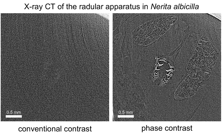
Phase contrast can also be combined with Computed Tomography (CT), i.e., 3D imaging. The image pair below shows the potential improvement phase contrast can achieve in challenging imaging scenarios. A gastropod of the family Nerita is imaged with its hard calcified shell. The radula, i.e., the "teeth" of the snail, is almost invisible in conventional CT, but with phase contrast it can be reproduced in great detail.

Reference
The first two images are from I. Häggmark, "Phase-Contrast X-ray Imaging of Complex Objects", doctoral thesis, KTH Royal Institute of Technology, Stockholm (2021). link to full-text
The CT images are from I. Häggmark, M. Hoshino et al., X-ray phase contrast reveals soft tissue and shell growth lines in mollusks, Communications Biology 7, 17 (2024). link to full-text